

 Current location: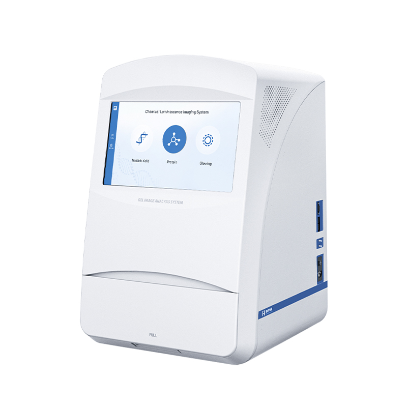
 +86
17853698681
+86
17853698681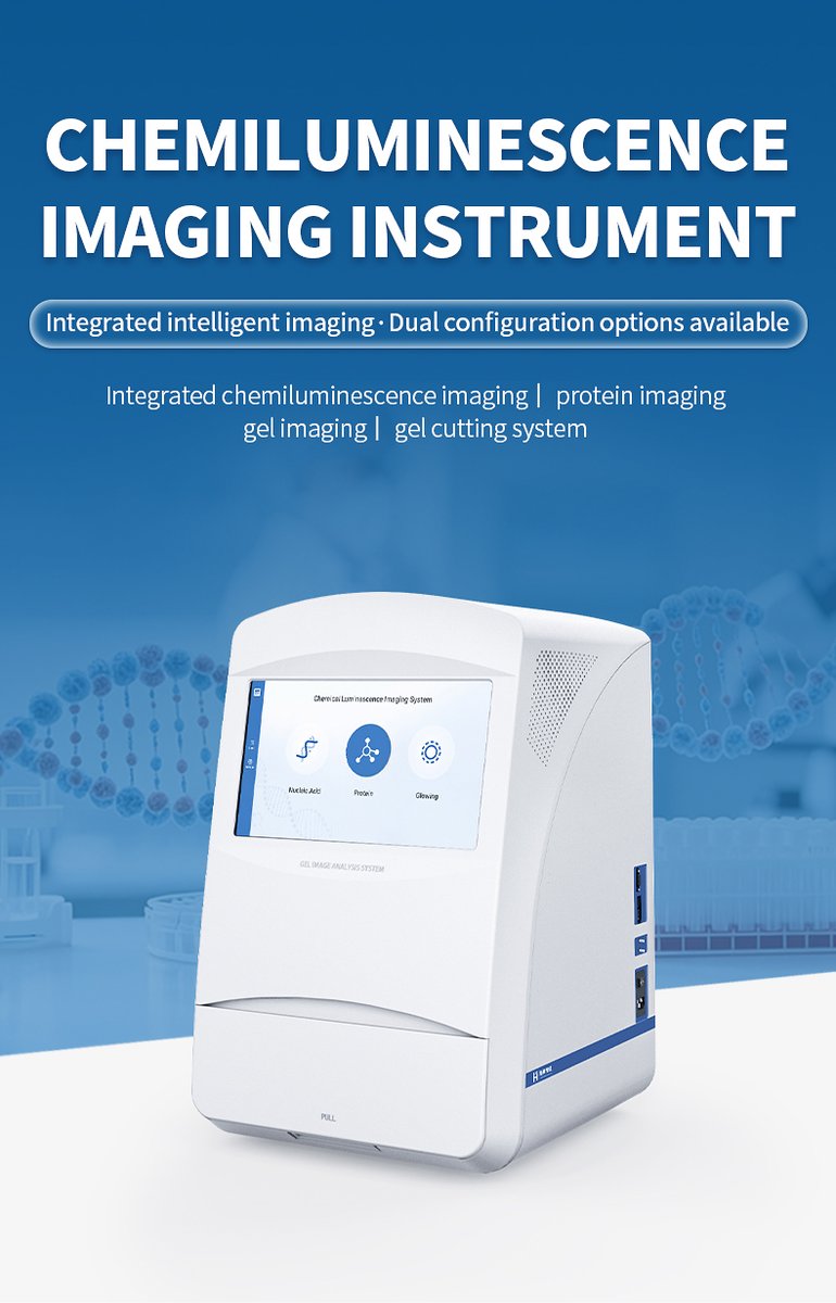


Product Introduction
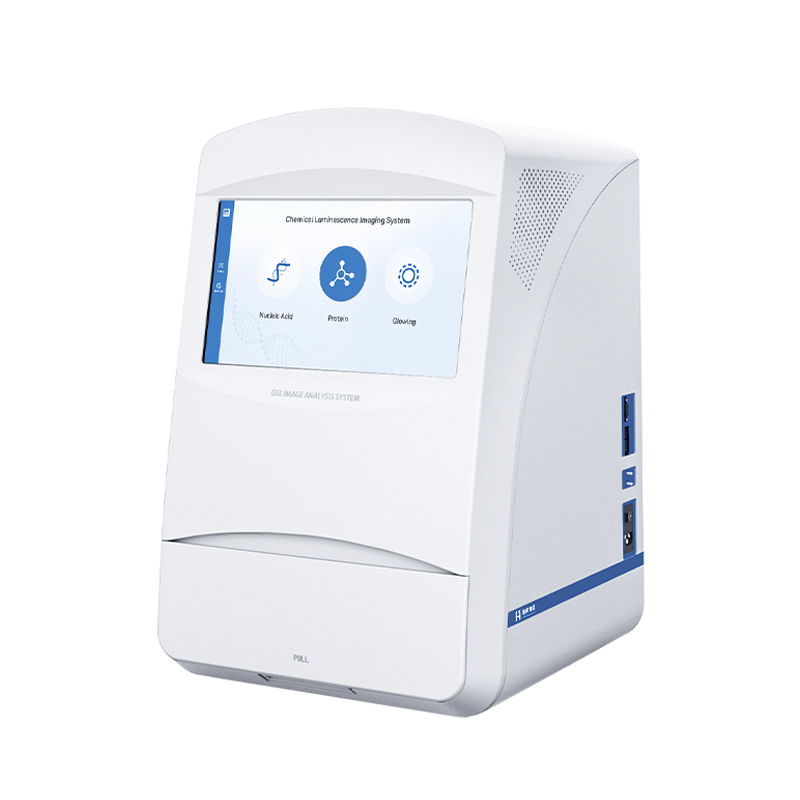
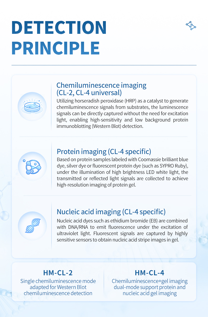
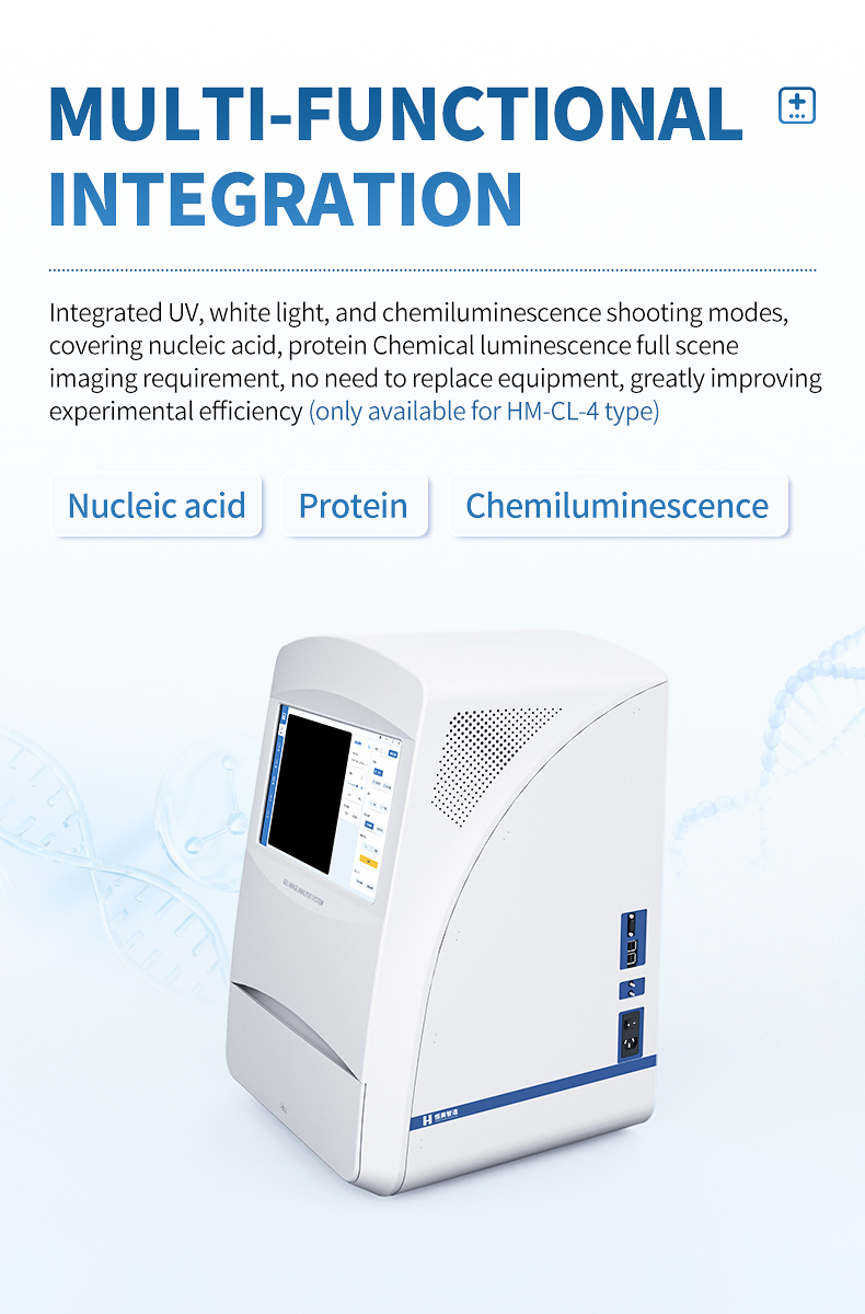
Molecular biology labs routinely handle two distinct imaging tasks: detecting proteins on Western Blot membranes and documenting nucleic acid gels. Traditionally, these tasks require separate instruments—a chemiluminescence imager for blots and a gel documentation system for DNA/RNA gels. The HM-CL-4 integrates both capabilities into a single benchtop platform, simplifying equipment management and reducing total cost of ownership.
The system's cooled CMOS sensor operates at -25 °C, delivering the low-noise performance required for long-exposure chemiluminescence. When switched to gel documentation mode, a 302 nm UV transilluminator and white-light LED strip provide excitation for fluorescent stains and colorimetric gels. The same F0.95 lens and 590 nm filter are shared across modes, maintaining consistent optical quality.
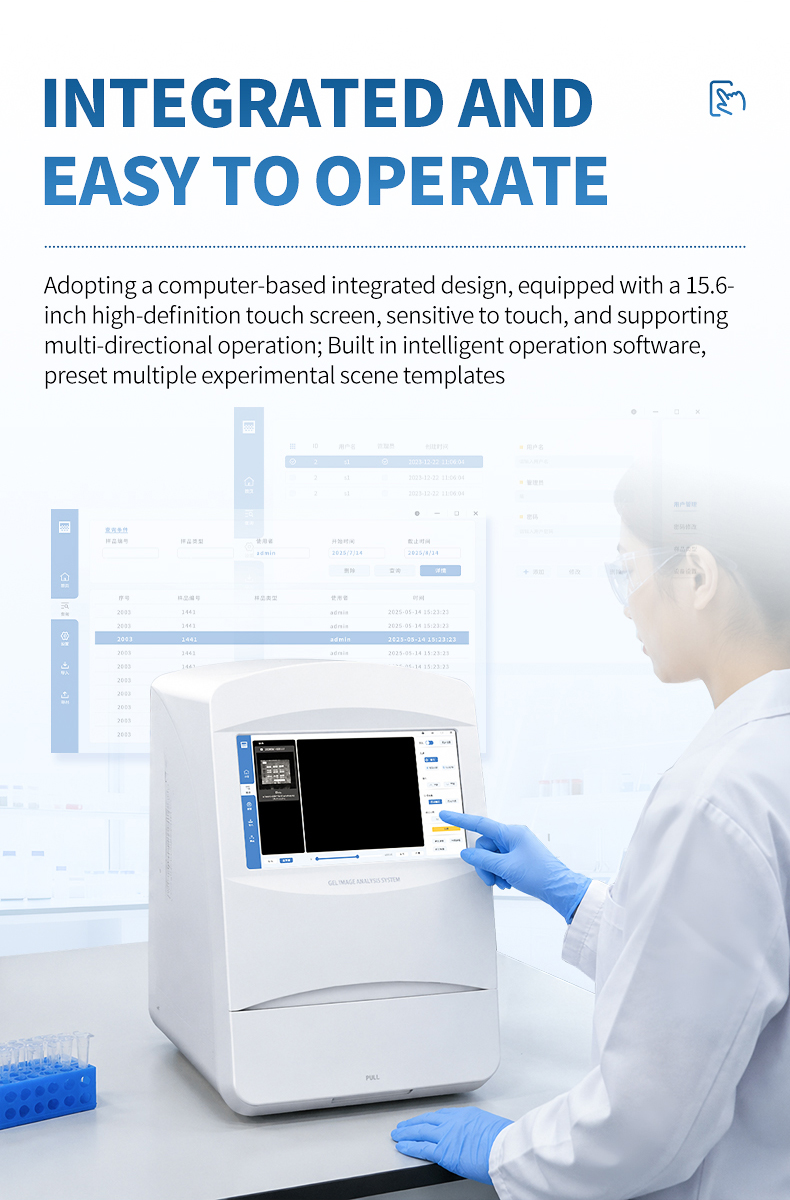
Researchers can document an agarose gel in the morning and capture a low-abundance phosphoprotein blot in the afternoon using the same instrument. Preset templates in the 15.6-inch touchscreen software guide users through each workflow, reducing training burden and minimizing protocol errors. The compact 460 × 470 × 715 mm footprint preserves valuable bench space in busy labs.
For labs that need a dedicated chemiluminescence-only system, the HM-CL-2 offers the same sensor and optics without the gel documentation hardware. For labs that want one instrument to cover both protein and nucleic acid imaging, the HM-CL-4 is the more efficient choice.
Applications
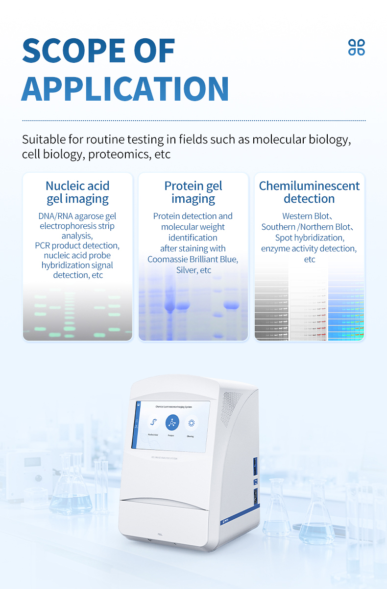
- Quantitative Western Blot: Relative and absolute protein expression analysis across treatment groups.
- Nucleic acid gel documentation: UV and white-light imaging of agarose and PAGE gels.
- Cloning verification: Rapid confirmation of PCR products and restriction digests.
- Antibody validation: Specificity and titer optimization for primary and secondary antibodies.
- Post-translational modification studies: Phosphorylation, acetylation, and ubiquitination detection.
- Reporter gene assays: Luciferase and enzyme activity measurements.
- Biopharma QC: Batch consistency, clone screening, and stability testing.
Key Features & Advantages
- Dual-mode design: Chemiluminescence + UV/white-light gel documentation in one system.
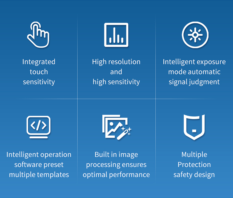
- No external computer: 15.6-inch integrated touchscreen controls all capture modes.
- Deep-cooled CMOS sensor: -25 °C cooling minimizes dark current for weak signals.
- 16-bit quantization: 65,536 gray levels for accurate densitometry.
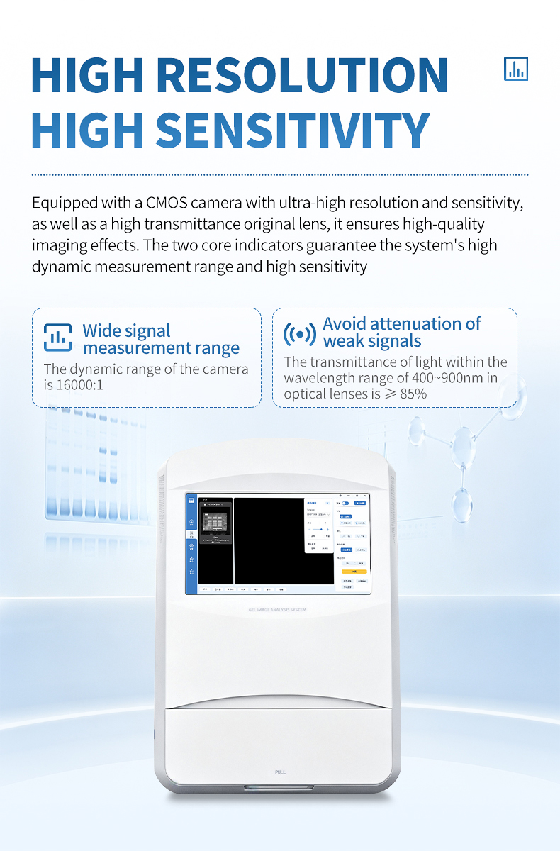
- Wide dynamic range: 16,000:1 captures faint and intense bands in one exposure.
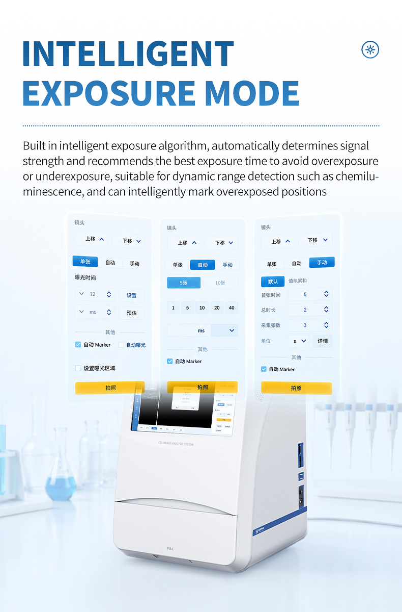
- Smart exposure: Algorithm recommends exposure time and flags saturated pixels.
- 590 nm narrow-band filter: Optimized emission filtering for chemiluminescence.
- 302 nm UV transilluminator: 6–10 W source with 3000 h life for nucleic acid gels.
- High-transmittance optics: F0.95 lens preserves weak signal intensity.
- Flexible binning: 1×1 through 8×8 adapts resolution and sensitivity.
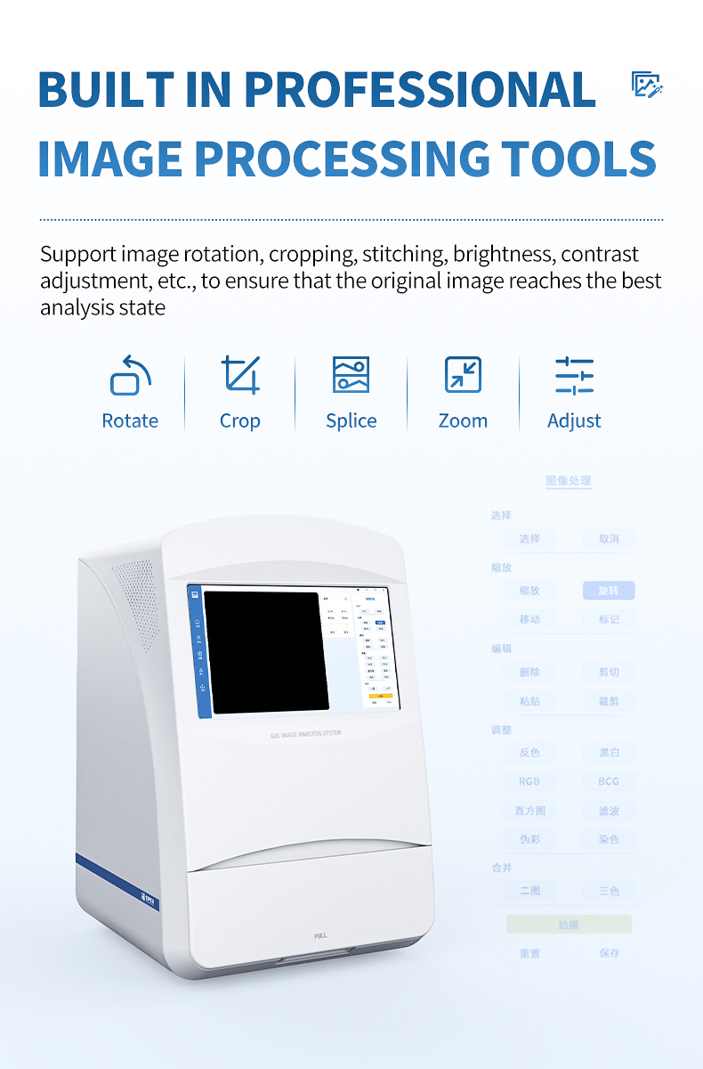
- Built-in image processing: Rotation, cropping, stitching, and contrast adjustment.
- Multiple export options: USB 2.0, USB 3.0, and Ethernet interfaces.
- Compact footprint: Fits standard lab benches at 460 × 470 × 715 mm.
- Low operating power: ≤ 90 W consumption reduces heat load.
Technical Specifications
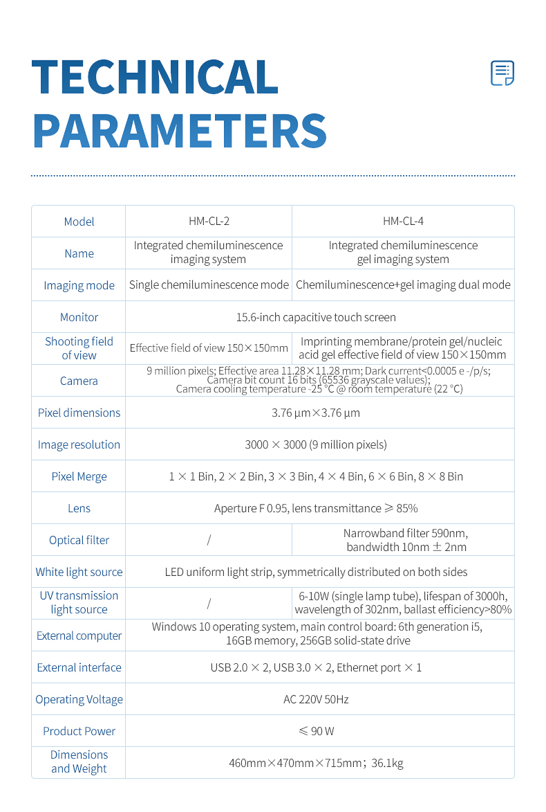
| Item | Specification |
|---|---|
| Display | 15.6-inch capacitive touchscreen |
| Camera | 9 MP cooled CMOS, 16-bit |
| Sensor cooling | -25 °C at 22 °C ambient |
| Dark current | < 0.0005 e-/p/s |
| Pixel size | 3.76 μm × 3.76 μm |
| Image resolution | 3000 × 3000 |
| Dynamic range | 16,000:1 |
| Imaging area | 150 mm × 150 mm effective field of view |
| Lens | F0.95, ≥ 85% transmittance at 400–900 nm |
| Filter | 590 nm narrow-band, bandwidth 10 nm ± 2 nm |
| Binning | 1×1, 2×2, 3×3, 4×4, 6×6, 8×8 |
| UV source | 302 nm transilluminator, 6–10 W, 3000 h life |
| White light source | LED uniform light strip |
| Interfaces | USB 2.0 × 2, USB 3.0 × 2, Ethernet × 1 |
| Operating voltage | AC 220V 50Hz |
| Power | ≤ 90 W |
| Dimensions | 460 × 470 × 715 mm |
| Weight | 36.1 kg |
FAQ
Q1: Can the HM-CL-4 replace both a Western Blot imager and a gel doc system?
Yes. The integrated UV/white-light source and chemiluminescence module cover nucleic acid gels, protein gels, and Western Blot membranes in one instrument.
Q2: Does adding gel documentation compromise chemiluminescence sensitivity?
No. The HM-CL-4 uses the same cooled CMOS sensor, F0.95 lens, and 590 nm filter as the HM-CL-2, so chemiluminescence performance is preserved.
Q3: Which fluorescent dyes are compatible with the UV transilluminator?
The 302 nm wavelength is suitable for ethidium bromide, SYBR Safe, SYBR Green, and other common nucleic acid stains excited near 300 nm.
Q4: How quickly can users switch between imaging modes?
Mode selection is done through the touchscreen software. Hardware switching is automatic, allowing mode changes in seconds without manual filter or lens swaps.
Q5: Is the system suitable for low-abundance phosphoprotein detection?
Yes. The combination of -25 °C cooling, 16-bit depth, and F0.95 optics supports detection of low-abundance targets with optimized antibodies and substrates.
Q6: Can images be exported for analysis in ImageJ or similar software?
Yes. Standard image files are exported via USB or Ethernet and can be opened in ImageJ, Quantity One, and other densitometry tools.
Q7: What maintenance is required for the UV lamp?
The 302 nm lamp has a rated life of 3000 hours. Users should clean the transilluminator surface regularly and track lamp hours to schedule replacement.
Article address:https://www.molecularbio.cn/chemiluminescence-imaging-system/molecular-biology-gel-imager-hm-cl4.html